วันอังคารที่ 29 พฤศจิกายน พ.ศ. 2554

อุปกรณ์ทางการแพทย์

หลอดเก็บเลือด ( Vacutainer )

 Containers containing anticoagulants

· Green - Contains Sodium Heparin or Lithium Heparin used for plasma determinations in clinical chemistry (e.g. urea and electrolyte determination).
· Light Green or Green/Gray 'Tiger': For plasma determinations.
· Purple or lavender - contains EDTA (the potassium salt, or K2EDTA). This is a strong anticoagulant and these tubes are usually used for full blood counts (CBC) and blood films. Lavender top tubes are generally used when whole blood is needed for analysis. Can also be used for some blood bank procedures such as blood type and screen, but other blood bank procedures, such as crossmatches must be in a pink tube in most facilities.
· Grey - These tubes contain fluoride and oxalate. Fluoride prevents enzymes in the blood from working, so a substrate such as glucose will not be gradually used up during storage. Oxalate is an anticoagulant.
· Light blue - Contain a measured amount of citrate. Citrate is a reversible anticoagulant, and these tubes are used for coagulation assays. Because the liquid citrate dilutes the blood, it is important the tube is full so the dilution is properly accounted for.
· Dark Blue - Contains sodium heparin, an anticoagulant. Also can contain EDTA as an additive or have no additive. These tubes are used for trace metal analysis.
· Pink - Similar to purple tubes (both contain EDTA) these are used for blood banking.
· Black - used for ESR also knows as Erythrocyte Sedimentation Rate or "sed rate." goes to hematology department.
ภาชนะบรรจุที่มี anticoagulants
สีเขียว -- ประกอบด้วยโซเดียมหรือเฮปารินเฮปารินลิเธียมที่ใช้สำหรับการวัดพลาสม่าในวิชาเคมีคลินิก (เช่นยูเรียและการกำหนดอิ)
สีเขียวอ่อนหรือสีเขียว / สีเทา
'ไทเกอร์': สำหรับการหาความพลาสม่า
ม่วงหรือดอกลาเวนเดอร์ -- มี
EDTA (เกลือโพแทสเซียมหรือ K2EDTA) นี้เป็นสารกันเลือดแข็งที่แข็งแกร่งและท่อเหล่านี้มักจะใช้สำหรับการนับจำนวนเลือดเต็มรูปแบบ (CBC) และฟิล์มเลือด ท่อด้านบนลาเวนเดอร์โดยทั่วไปจะใช้เมื่อเลือดเป็นสิ่งจำเป็นสำหรับการวิเคราะห์ นอกจากนี้ยังสามารถใช้สำหรับการบางขั้นตอนการธนาคารเลือดเช่นชนิดของเลือดและหน้าจอ แต่วิธีการที่ธนาคารเลือดอื่น ๆ เช่น crossmatches จะต้องอยู่ในท่อสีชมพูในสิ่งอำนวยความสะดวกมากที่สุด
สีเทา -- หลอดเหล่านี้มีฟลูออไรและออกซาเลต ลูออไรด์ป้องกันไม่ให้เอนไซม์ในเลือดจากการทำงานเพื่อให้พื้นผิวเช่นน้ำตาลกลูโคสที่จะไม่ถูกใช้ค่อยๆขึ้นระหว่างการเก็บรักษา ออกซาเลตเป็นสารกันเลือดแข็ง
ฟ้า -- มีจำนวนเงินที่วัดของ
citrate citrate เป็น anticoagulant ย้อนกลับและท่อเหล่านี้จะใช้สำหรับการสอบตรวจการแข็งตัว เนื่องจากซิเตรทเจือจางของเหลวในเลือดเป็นสิ่งสำคัญที่หลอดเป็นอย่างเต็มรูปแบบเพื่อการลดสัดส่วนที่จะคิดอย่างถูกต้องสำหรับ
Dark Blue -- ประกอบด้วยโซเดียมเฮ, anticoagulant ยังสามารถมี EDTA เป็นสารเติมแต่งหรือมีสารที่ไม่มี หลอดเหล่านี้จะใช้สำหรับการวิเคราะห์โลหะติดตาม
ชมพู -- คล้ายกับหลอดสีม่วง (ทั้งสองมี
EDTA) เหล่านี้จะใช้สำหรับการธนาคารเลือด
สีดำ -- ใช้สำหรับการ
ESR ยังรู้อัตราการตกตะกอนของเม็ดเลือดแดงเป็นหรือ"อัตรา sed." ไปที่แผนกโลหิตวิทยา

วันจันทร์ที่ 21 พฤศจิกายน พ.ศ. 2554

Definition

Definition
 1. A pneumonia is bacteria disease caused which  infection the cause bronchial pneumonia and  acute  interstitial pneumonia.
 2. A cirrhosis is disease causes which  chronic infection of hepatitis virus B or C  will have The symptoms Malaise .
 3. insomnia is caused stress This is often a symptom of anxiety disorders or the delayed sleep phase disorder.

 4. A anorexia  which the symptom of poor appetite whatever the cause Anorexia .
 5. immunity  is which of an organism of  as an infection or disease.
 6. intense itching and superficial burrows is called a scabies.
 7. Colles' fracture which distal radius in the forearm with dorsal  displacement of the wrist  sometimes and hand is called  a "dinner fork.
8. peptic ulcer is sores on the birth of the lining of the stomach or small intestine which has abdominal pain before or after eating food at all times and very often do not detect anything unusual.
9. Encephalitis means inflammation of the brain which has
The inflammation may affect the brain or part of the brain. The cause of encephalitis is usually caused by a virus related.
10. Thyroid hormone that is released into the body the metabolism of nutrients, too will cause the symptoms is called  toxic thyroid (Hyperthyroidism) or toxic goiter.

วันพุธที่ 16 พฤศจิกายน พ.ศ. 2554

Narration (การบรรยายหรือการเล่าเรื่อง)

Narration
This paragraph pattern tells a story about a series of event or actions. The story can be fiction (not based on facts) or it can be nonfiction (based on real-life experiences). Signaled by such phrases as, in the beginning, at first, afterwards, in conclusion, and to sum up. Sometimes, you can see the grammar in the paragraph like this. : story >> past tense, history >> present perfect, beginning >> past perfect and ending >> adverb of time
เป็นการเล่าเรื่องราวที่มีหลักเกณฑ์ในการเล่าเรื่อง 2 ประเภท
  1. บรรยายการเล่าเรื่องราวโดยอาศัยหลักการความเป็นจริงหรือเหตุการณ์ที่เกิดขึ้นจริงโดยนำมาเล่าเป็นการบรรยาย เช่น วรรณคดี วรรณกรรม หรือประสบการณ์ที่เกี่ยวกับผู้อื่น
  2. ประเภทที่ไม่อาศัยข้อเท็จจริงเป็นเรื่องราวหรือการบรรยายที่แต่งขึ้นเอง เช่น นวนิยาย  นิทาน  เรื่องตลกขบขัน
ตัวอย่างเช่น นิทานเรื่อง
The Elephant’s Child
based on Just So Stories by Rudyard Kipling
by Logan Gardner, age 6
The elephant wanted to know what crocodiles eat for supper. So the elephant went to the river to see what they eat.
ช้างต้องการที่จะรู้ว่าสิ่งที่จระเข้กินสำหรับอาหารเมื้อเย็นคืออะไร ดั้งนั้น  ช้างจึงไปที่แม่น้ำเพื่อจะเห็นสิ่งที่จระเข้กิน
The crocodile winked with one eye and then with the other eye. The elephant said, “I just want to know what crocodiles eat.”
จระเข้จ่องช้างด้วยหน้าตาที่หน้ากลัว ช้างจึงกล่าวว่า ผมเพียงต้องการที่จะรู้ว่าสิ่งที่จระเข้กินคืออะไร
The crocodile said, “Come here and I’ll tell you.”
จระเข้บอกว่า มาที่นี้แล้วฉันจะบอกคุณ
The elephant got away. Then the elephant went home to his mother and father , brothers and sisters. He kissed them all.
ช้างก็กลับบ้านไปหาพ่อแม่และพี่น้องของเขา เขาจูบพวกเขาทั้งหมด
The elephant got close. Then the crocodile bit his nose. The crocodile pulled. And the elephant pulled the other way. The elephant’s nose got longer and longer when they both pulled.
ช้างจึงเข้าไปอย่างชิด จากนั้นจระเข้จึงบิดจมูกของเขา จระเข้ดึงและช้างก็ดึงจมูกของเขากลับมาด้วยวิธีการอื่นๆ จนสามารถดึงจมูกของเขากลับมาคืนได้

วันอังคารที่ 15 พฤศจิกายน พ.ศ. 2554

Encephalitis

Encephalitis


Encephalitis is an acute inflammation of the brain. Encephalitis with meningitis is known as meningoencephalitis. Symptoms include headache, fever, confusion, drowsiness, and fatigue. More advanced and serious symptoms include seizures or convulsions, tremors, hallucinations, and memory problems.

Cause

Viral

Main article: Viral encephalitis
Viral encephalitis can occur either as a direct effect of an acute infection, or as one of the sequelae of a latent infection. The most common causes of viral encephalitis are Rabies, Herpies Simplex, Polio, SSPE and PML.[1] Other causes include infection by flaviviruses such as St. Louis encephalitis or West Nile virus, or by Togaviridae such as Eastern equine encephalitis (EEE), Western equine encephalitis (WEE) or Venezuelan equine encephalitis (VEE).

 Bacterial and other

It can be caused by a bacterial infection, such as bacterial meningitis, spreading directly to the brain (primary encephalitis), or may be a complication of a current infectious disease syphilis (secondary encephalitis). Certain parasitic or protozoal infestations, such as toxoplasmosis, malaria, or primary amoebic meningoencephalitis, can also cause encephalitis in people with compromised immune systems. Lyme disease and/or Bartonella henselae may also cause encephalitis.
Another cause is granulomatous amoebic encephalitis.

 Diagnosis

Adult patients with encephalitis present with acute onset of fever, headache, confusion, and sometimes seizures. Younger children or infants may present irritability, poor appetite and fever.
Neurological examinations usually reveal a drowsy or confused patient. Stiff neck, due to the irritation of the meninges covering the brain, indicates that the patient has either meningitis or meningoencephalitis. Examination of the cerebrospinal fluid obtained by a lumbar puncture procedure usually reveals increased amounts of protein and white blood cells with normal glucose, though in a significant percentage of patients, the cerebrospinal fluid may be normal. CT scan often is not helpful, as cerebral abscess is uncommon. Cerebral abscess is more common in patients with meningitis than encephalitis. Bleeding is also uncommon except in patients with herpes simplex type 1 encephalitis. Magnetic resonance imaging offers better resolution. In patients with herpes simplex encephalitis, electroencephalograph may show sharp waves in one or both of the temporal lobes. Lumbar puncture procedure is performed only after the possibility of prominent brain swelling is excluded by a CT scan examination. Diagnosis is often made with detection of antibodies in the cerebrospinal fluid against a specific viral agent (such as herpes simplex virus) or by polymerase chain reaction that amplifies the RNA or DNA of the virus responsible (such as varicella zoster virus).

 Treatmen

Treatment is usually symptomatic. Reliably tested specific antiviral agents are few in number (e.g. acyclovir for herpes simplex virus) and are used with limited success in treatment of viral infection, with the exception of herpes simplex encephalitis. In patients who are very sick, supportive treatment, such as mechanical ventilation, is equally important. Corticosteroids (e.g. methylprednisolone) are used to reduce brain swelling and inflammation. Sedatives may be needed for irritability or restlessness.

Encephalitis lethargica

Encephalitis lethargica is an atypical form of encephalitis which caused an epidemic from 1918 to 1930. Those who survived sank into a semi-conscious state that lasted for decades until the Parkinson's drug L-DOPA was used to revive those still alive in the late 1960s by Oliver Sacks.
There have been only a small number of isolated cases in the years since, though in recent years a few patients have shown very similar symptoms. The cause is now thought to be either a bacterial agent or an autoimmune response following infection.
Encephalitis หมายถึงมีการอักเสบของเนื้อสมอง
การอักเสบอาจจะเกิดทั้งสมองหรือบางส่วนของเนื้อสมอง สาเหตุของสมองอักเสบมักจะเกิดจากไวรัสเช่น
เริม คางทูม หัดเยอรมัน ไข้สุกใส นอกจากนั้นยังเกิดจากยุงหรือไรกัดเช่น Japanese encephalitis
อาการของผู้ป่วยสมองอักเสบ ผู้เป็นไม่มากจะมีอาการ
ไข้
อ่อนเพลียไม่มีแรง
เจ็บคอ
คอแข็ง
อาเจียน
ปวดศีรษะ
สับสน
กระสับกระส่าย
ซึม
ปวดหัวเมื่อแสงจ้าๆ 


สำหรับผู้ที่มีอาการมากได้แก่
ซึม ไม่ค่อยรู้สึกตัว
สับสนไม่รู้วันหรือกลางคืน จำคนไม่ได้
ชัก
ไข้สูง
ปวดศีรษะมาก
คลื่นไส้อาเจียน
มือสั่น
คอแข็ง
สาเหตุ สาเหตุของสมองอักเสบส่วนใหญ่เกิดจากเชื้อไวรัส การติดต่อโดยมากเกิดจากยุง ไรกัด เช่นโรคสมองอักเสบจากไวรัสสายพันธ์ยี่ปุ่น japaness encephalitis บางชนิดเกิดจากการที่สัตว์เช่นค้างคาวหรือสุนัขกัด
 เช่นโรคหมาบ้า
ประเภทของการติดเชื้อแบ่งออกเป็น 2 ประเภทคือ
*ติดเชื้อครั้งแรก Primary encephalitis หมายถึงการติดเชื้อไวรัสเป็นครั้งแรกและเชื้อนั้นก็ทำให้เกิดสมองอักเสบ
มักจะมีการระบาดเป็นครั้งคราว เช่นไขสมองอักเสบสายพันธ์ญี่ปุ่น
*สมองอักเสบซึ่งเกิดจากเชื้อที่อยู่ในร่างกายเรียก Secondary (post-infectious) encephalitis เช่นสมองอักเสบจากเชื้อเริม

Myxedema

Myxedema

Myxedema (British English: myxoedema) describes a specific form of cutaneous and dermal edema secondary to increased deposition of connective tissue components (like glycosaminoglycans, hyaluronic acid, and other mucopolysaccharides) in subcutaneous tissue as seen in various forms of hypothyroidism and Graves' disease.[1]:535 It is more common in women than in men. [2]

Terminology

The word originates from μύξα, taken from ancient Greek to convey 'mucus' or 'slimy substance' and ίδημα for swelling.
Myxedema coma is rare and establishing the diagnosis requires a high index of suspicion. Myxedema coma represents the severest form of hypothyroidism and has an associated mortality rate of 30 percent to 40 percent. It can occur due to long-standing, untreated hypothyroidism, but is often linked to a precipitant, such as acute infection, myocardial infarction, congestive heart failure, cerebral vascular accident, trauma, or drug toxicity. Several medications can cause hypothyroidism, and patients taking them must be carefully monitored. These medications include amiodarone, lithium, and sedatives. No consensus exists on specific thyroid hormone replacement regimens for myxedema coma. Most experts agree that a large intravenous bolus of levothyroxine should be administered (200 to 400 mcg), followed by daily doses of 50 to 100 mcg, based on the patient's weight and comorbidities. Other experts advocate the use of triiodothyronine (T3) or a combination of both T3 and T4. In addition to thyroid replacement therapy, it is important to detect coexisting adrenal insufficiency and treat patients with stress-dose steroids to avoid precipitating adrenal crisis.[5]

 Cause

The increased deposition of glycosaminoglycan is not fully understood, however two mechanisms predominate.
  • Exophthalmos in particular results from TSH receptor stimulation on fibroblasts behind the eyes which leads to increased glycosaminoglycan deposition. It is thought that many cells responsible for forming connective tissue react to increases in TSH levels.[citation needed]
  • Secondarily, in autoimmune thyroid diseases lymphocytes react to the TSH receptor. Thus, in addition to the inflammation within the thyroid, any cell that expresses the TSH receptor will likely experience lymphocytic infiltrates as well. The inflammation can cause tissue damage and scar tissue formation, explaining the deposition of glycosaminoglycans.[citationeeded
The increased deposition of glycosaminoglycans causes an osmotic edema and fluid collection.
Hashimoto's thyroiditis is the most common cause of myxedema in the United States.[6]
Myxedema (อังกฤษ : myxoedema) อธิบายรูปแบบเฉพาะของอาการบวมน้ำและผิวหนังผิวหนังรองลงไปสะสมที่เพิ่มขึ้นขององค์ประกอบของเนื้อเยื่อเกี่ยวพัน (เช่น glycosaminoglycans, กรดไฮยาลูโร, และ mucopolysaccharides อื่น ๆ ) ในเนื้อเยื่อใต้ผิวหนังตามที่เห็นในรูปแบบต่างๆของ hypothyroidism และโรค Graves' [1] : 535 มันเป็นพบมากในผู้หญิงมากกว่าในผู้ชาย [2]
สาเหตุ
เกิดการสะสมที่เพิ่มขึ้นของ glycosaminoglycan; คือไม่เข้าใจอย่างเต็มที่ แต่มีอิทธิพลเหนือกลไกทั้งสอง
Exophthalmos ในผลโดยเฉพาะอย่างยิ่งจากการกระตุ้นตัวรับบน fibroblasts TSH หลังตาซึ่งนำไปสู่​​การสะสมเพิ่มขึ้น glycosaminoglycan; มันเป็นความคิดที่หลายเซลล์ที่รับผิดชอบในการสร้างเนื้อเยื่อเกี่ยวพันตอบสนองต่อการเพิ่มขึ้นในระดับ TSH. [อ้างอิงที่จำเป็น]
ครั้งที่สองในโรคต่อมไทรอยด์ภูมิต้านทานเนื้อเยื่อเซลล์เม็ดเลือดขาวทำปฏิกิริยากับตัวรับ
TSH ดังนั้นนอกเหนือไปจากการอักเสบของต่อมไทรอยด์ภายในเซลล์ที่แสดงตัวรับ TSH ใด ๆ มีแนวโน้มที่จะแทรกตัวเข้าไปใน lymphocytic ประสบการณ์เป็นอย่างดี การอักเสบอาจทำให้เกิดการทำลายเนื้อเยื่อและการสร้างเนื้อเยื่อแผลเป็นอธิบายการสะสมของ glycosaminoglycans. [citationeeded
สะสมที่เพิ่มขึ้นของ
glycosaminoglycans ทำให้เกิดอาการบวมน้ำที่ออสโมติกและการเก็บรวบรวมของเหล
Hashimoto ของ thyroiditis เป็นสาเหตุที่พบมากที่สุดของ Myxedema ในประเทศสหรัฐอเมริกา. [6]


Colles' fracture

Colles' fracture


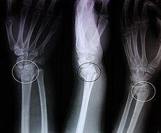
A Colles' fracture, also Colles fracture, is a fracture of the distal radius in the forearm with dorsal (posterior) displacement of the wrist and hand. The fracture is sometimes referred to as a "dinner fork" or "bayonet" deformity due to the shape of the resultant forearm. For a more detailed discussion see distal radius fracture.

Terminology

The term Colles fracture is classically used to describe a fracture at the distal end of the radius, at its cortico-cancellous junction. However, now the term tends to be used loosely to describe any fracture of the distal radius, with or without involvement of the ulna, that has dorsal displacement of the fracture fragments. Colles himself described it as a fracture that “takes place at about an inch and a half (38mm) above the carpal extremity of the radius” and “the carpus and the base of metacarpus appears to be thrown backward”.[2]
The classic Colles fracture has the following characteristics:[3]

Causes

The fracture is most commonly caused by people falling onto a hard surface and breaking their fall with outstretched arms - falling with wrists flexed would lead to a Smith's fracture. It can also be caused by overuse. Orignally it was described in elderly people, post-menopausal women. It usually occurs about an inch or two proximal to the radio-carpal joint with posterior and lateral displacement of the distal fragment resulting in the characteristic "dinner fork" or "bayonet" like deformity.
Colles' fracture is a common fracture in people with osteoporosis, second only to vertebral fractures.

Diagnosis

  • Dorsal tilt
  • Radial shortening
  • Loss of ulnar inclination
  • Radial angulation of the wrist
  • Dorsal displacement of the distal fragment
  • Comminution at the fracture site
  • Associated fracture of the ulnar styloid process in more than 60% of cases.

Treatment

Management depends on the severity of the fracture. An undisplaced fracture may be treated with a cast alone.The cast is applied with the distal fragment in palmar flexion and ulnar deviation. A fracture with mild angulation and displacement may require closed reduction. There is some evidence that immobilization with the wrist in dorsiflexion as opposed to palmarflexion results in less redisplacement and better functional status.[4] Significant angulation and deformity may require an open reduction and internal fixation or external fixation. The volar forearm splint is best for temporary immobilization of forearm, wrist and hand fractures, including Colles' fracture There are several established instability criteria: dorsal tilt >20°, comminuted fracture, abruption of the ulnar styloid process, intraarticular displacement >1mm, loss of radial height >2mm.
A higher amount of instability criteria increases the likelihood of operative treatment.
Treatment modalities differ in the elderly.[5]

Eponym

t is named after Abraham Colles (1773–1843), an Irish surgeon who first described it in 1814 just by looking at the classical deformity before the advent of X-rays.[6] Ernest Amory Codman was the first to study it using X-rays. His article, published in the Boston Medical and Surgical Journal, now known as The New England Journal of Medicine, also developed the classification system.[7][8]

In older and younger people

Colles fractures occur in all age groups, although certain patterns follow an age distribution.
  • In the elderly, because of the weaker cortex, the fracture is more often extra-articular.
  • Younger individuals tend to require a higher energy force to cause the fracture and tend to have more complex intra-articular fractures. In children with open epiphyses, an equivalent fracture is the "epiphyseal slip", as can be seen in other joints, such as a slipped capital femoral epiphysis in the hip. This is a Salter I or II fracture with the deforming forces directed through the weaker epiphyseal plate.
  • More common in women because of post-menopausal osteoporosis.

References

  ^ Al-Ansari K, Howard A, Seeto B, Yoo S, Zaki S, Boutis K (2007). "Minimally angulated pediatric wrist fractures: is immobilization without manipulation enough?". CJEM : Canadian journal of emergency medical care = JCMU : journal canadien de soins médicaux d'urgence 9 (1): 9–15. PMID 17391594.[dead link]
 ^ Colles A 2006 On the fracture of the carpal extremity of the radius. Edinb Med Surg J. 1814;10:181. Clin Orthop Relat Res 445:5-7.
  ^ GP Notebook. "Colles' fracture". Retrieved 2009-02-21.
  ^ William G Blakeney.Stabilization and treatment of Colles’ fractures in elderly patients. http://www.dovepress.com/articles.php?article_id=5677
  ^ Mallon, Bill (2000). Ernest Amory Codman : the end result of a life in medicine. Philadelphia: Saunders. ISBN 9780721684611.
  ^ CODMAN, E. A. (1900). "A Study of the X-Ray Plates of One Hundred and Forty Cases of Fracture of the Lower End of the Radius". The Boston Medical and Surgical Journal 143 (13): 305–308. doi:10.1056/NEJM190009271431301. ISSN 0096-6762.

การแตกหัก Colles
สาเหตุการแตกหักจะเกิดขึ้นมากที่สุดโดยคนที่ล้มลงบนพื้นผิวที่แข็ง จะนำไปสู่​​การแตกหัก Smith ของ กระดูก นอกจากนี้ยังสามารถเกิดจากการใช้มากเกินไป ในผู้สูงอายุสตรีวัยหมดประจำเดือนโพสต์ มักจะเกิดขึ้นเกี่ยวกับนิ้ว กับการกระจัดหลังและด้านข้างของส่วนปลาย
การแตกหัก
Colles'เป็นกระดูกที่พบบ่อยในคนที่มีโรคกระดูกพรุนที่สองเท่านั้นที่กระดูกพรุน
การรักษาการบริหารจัดการขึ้นอยู่กับความรุนแรงของการแตกหักที่การแตกหัก undisplaced อาจจะรักษาด้วยการโยนโยน alone.The ถูกนำไปใช้กับส่วนปลายในการงอ Palmar และ ulnar เบี่ยงเบน การแตกหักของกระดูกที่มี angulation อ่อนและผลอาจจำเป็นต้องลดการปิด มีหลักฐานบางอย่างที่ตรึงกับข้อมือใน dorsiflexion เป็นนอกคอกผล palmarflexion ใน redisplacement น้อยลงและสถานะการทำงานที่ดีกว่า. [4] angulation ที่สําคัญและความผิดปกติอาจต้องใช้การลดการเปิดและการตรึงภายในหรือตรึงภายนอก เข้าเฝือกแขน Volar ที่ดีที่สุดสำหรับการตรึงชั่วคราวของกระดูกหักที่ข้อมือแขนและมือรวมทั้งการแตกหัก Colles'มีหลายที่จัดตั้งขึ้นตามเกณฑ์ความไม่แน่นอน : เอียงหลัง> 20 °, แตกหัก abruption comminuted, ของกระบวนการ styloid ulnar, พลัดถิ่น intraarticular> 1mm, การสูญเสีย ความสูงของรัศมี> 2mm
จำนวนเงินที่สูงกว่าเกณฑ์ความไม่แน่นอนเพิ่มโอกาสในการรักษาผ่าตัด
การรักษาแบบวิธีที่แตกต่างกันในผู้สูงอายุ. [5]
ในผู้สูงอายุและเด็กกระดูกหัก Colles เกิดขึ้นในทุกกลุ่มอายุ แต่บางรูปแบบการกระจายตามอายุ     ในผู้สูงอายุเพราะของเยื่อหุ้มสมองลดลงของการแตกหักมักจะมีมากขึ้นเกี่ยวกับข้อ - พิเศษ     บุคคลที่อายุน้อยกว่ามีแนวโน้มที่จะต้องบังคับให้พลังงานที่สูงขึ้นจะก่อให้เกิดการแตกหักและมีแนวโน้มที่จะมีกระดูกหักภายในเกี่ยวกับข้อ - ซับซ้อนมากขึ้น ในเด็กที่มี epiphyses เปิดการแตกหักเทียบเท่าคือ"ใบ epiphyseal"เป็นสามารถเห็นได้ในข้อต่ออื่น ๆ เช่น epiphysis เล็ดรอดทุนในต้นขาสะโพก นี้เป็น Salter I หรือ II แตกหักกับกองกำลังเปลี่ยนรูปโดยตรงผ่านแผ่น epiphyseal ที่อ่อนลง พบมากในผู้หญิงเพราะของโรคกระดูกพรุนหลังวัยหมดประจำเดือน